Did you ever wonder what the inside of a heart looks like? Now that we have new technology, we can actually see what parts of the heart look like using an anatomy models 3d. Let’s explore this fascinating part of the body, the world of the heart and the processes that make it tick!
A 3D heart medical models can teach us a little about what the heart is. The heart is a vital organ that circulates blood throughout our body. It is what allows oxygen and nutrients to get to our cells. We can understand how this amazing organ keeps us healthy and alive by taking a tour through a 3D model of the heart.

When we look at a 3 D heart model, we see that the heart consists of four chambers, the left and right atria and the left and right ventricles. These chambers work together so that blood goes where it should. This Human model ensures that all of our body receives the nutrients, and oxygen it requires to function.
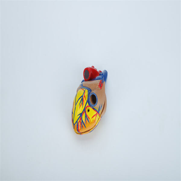
A 3D model of the heart provides a clearer view of the chambers and valves and allows us to see that the heart is also one of the strongest muscles in the body. The Valves of the heart are very important. They regulate the flow of blood, ensuring it’s all going in the right direction and that none of it is sloshing around backward. When we consider a Biological experiment kit 3D model of the heart, finding out how the valves are opening and closing at each beat, we are able to detect if the blood is flowing without obstructions.

Having a 3D model for how the heart functions is important for our heart health. By understanding how the heart functions in 3-D, we can see why it’s so important to keep it healthy with exercise, healthy eating and regular visits to the doctor. The heart is such an incredible organ that does the most amazing thing to keep us alive. We can appreciate its specialness by examining its parts with a 3D model.
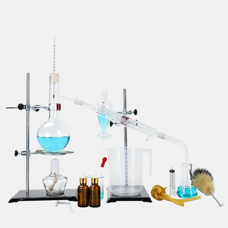
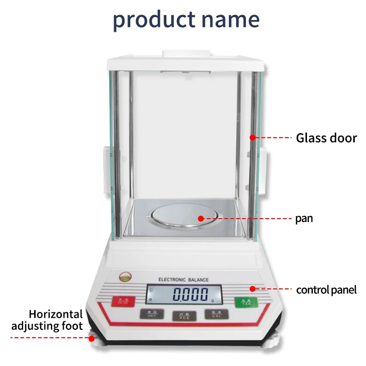
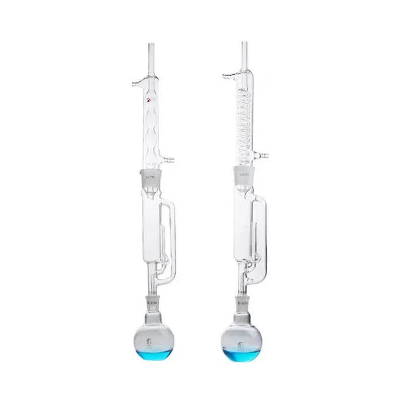
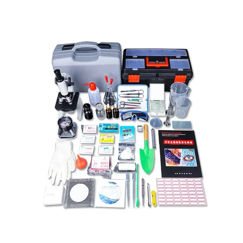
We offer a complete portfolio of science lab equipment across physics, chemistry, biology, and primary sciences, with a dedicated R&D team that provides tailored solutions for curriculum and laboratory needs.
Our products hold ISO 9001:2015, CE certifications, and test reports. We provide full lifecycle support—from custom design and safe packaging to warranty services and post-warranty repairs—ensuring a seamless customer experience.
With a team of over 20 foreign trade professionals, we export to 30+ regions including Europe, the U.S., the Middle East, Africa, and Asia, supported by fast production, efficient logistics, and timely delivery.
As a source manufacturer with 20 years of experience, we independently handle production, R&D, and global export, ensuring full quality control and reliable supply for educational equipment.
 EN
EN
 AR
AR
 BG
BG
 HR
HR
 CS
CS
 DA
DA
 NL
NL
 FI
FI
 FR
FR
 DE
DE
 EL
EL
 HI
HI
 IT
IT
 JA
JA
 KO
KO
 PL
PL
 PT
PT
 RO
RO
 RU
RU
 ES
ES
 TL
TL
 ID
ID
 SR
SR
 SK
SK
 SL
SL
 VI
VI
 MT
MT
 TH
TH
 TR
TR
 FA
FA
 AF
AF
 MS
MS
 GA
GA
 HY
HY
 BN
BN
 MN
MN
 SO
SO
 KK
KK
 MY
MY
 UZ
UZ